Polysaccharide Composites as a Wound-Healing Sponge
Ayesha Khalid, Naveera Naeem, Taous Khan and Fazli Wahid
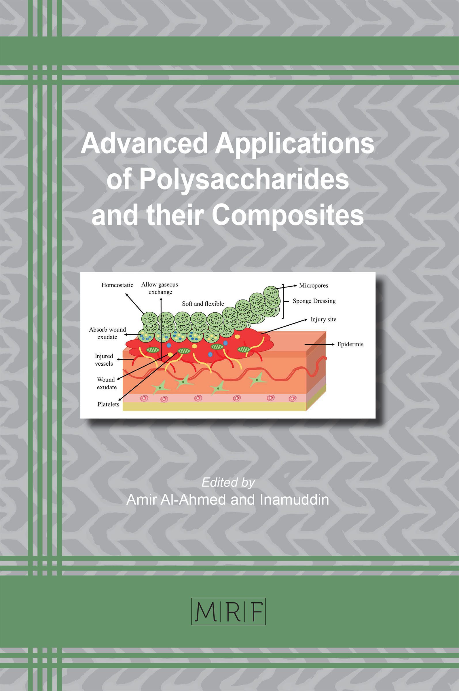
The first wound treatment was designed five millennia ago. Since, then tremendous scientific and technological advancements have been made to design multifunctional wound healing materials. This chapter gives a comprehensive discussion on the molecular and cellular events of wound repair and the biological aspects of polysaccharide-based sponges as a dressing system. Overall, this chapter focuses on the polysaccharides including alginate, cellulose, chitosan, hyaluronic acid, dextran and others. It briefly recaps the recent progress made in developing their composite sponges. Furthermore, the healing performance of these sponges is also discussed in detail.
Keywords
Wound Healing, Polysaccharide, Sponges, Dressing, Composites
Published online 4/20/2020, 26 pages
Citation: Ayesha Khalid, Naveera Naeem, Taous Khan and Fazli Wahid, Polysaccharide Composites as a Wound-Healing Sponge, Materials Research Foundations, Vol. 73, pp 1-26, 2020
DOI: https://doi.org/10.21741/9781644900772-1
Part of the book on Advanced Applications of Polysaccharides and their Composites
References
[1] E. Mclafferty, C. Hendry, A. Farley, The integumentary system: anatomy,physiology and function of skin, Nurs. Stand. 27 (2012) 35. https://doi.org/10.7748/ns2012.10.27.7.35.c9358
[2] L. Watret, R. White, Surgical wound management: the role of dressings. Nurs. Stand. 44 (2001) 59-69.
[3] B. M. Delavary, W. M. van der Veer, M. van Egmond, F. B. Niessen, R. H. Beelen, Macrophages in skin injury and repair, Immunobiology. 216 (2011) 753-762. https://doi.org/10.1016/j.imbio.2011.01.001
[4] P. Martin, Wound healing-aiming for perfect skin regeneration, Science. 276 (1997) 75-81. https://doi.org/10.1126/science.276.5309.75
[5] R. A. Clark, The molecular and cellular biology of wound repair, Ed Springer Science & Business Media, 2013.
[6] B. Behm, P. Babilas, M. Landthaler, S. Schreml, Cytokines, chemokines and growth factors in wound healing, J. Eur. Acad. Dermatol. Venereol. 26 (2012) 812-820. https://doi.org/10.1111/j.1468-3083.2011.04415.x
[7] S. Barrientos, O. Stojadinovic, M.S. Golinko, H. Brem, M. Tomic-Canic, Growth factors and cytokines in wound healing, Wound Repair Regen.16 (2008) 585-601. https://doi.org/10.1111/j.1524-475X.2008.00410.x
[8] T. Velnar, T. Bailey, V. Smrkolj, The wound healing process: an overview of the cellular and molecular mechanisms, J. Int. Med. Res. 37 (2009) 1528-1542. https://doi.org/10.1177/147323000903700531
[9] N. B. Menke, K. R. Ward, T. M. Witten, D. G. Bonchev, R. F. Diegelmann, Impaired wound healing, Clin. Dermatol. 25 (2007) 19-25. https://doi.org/10.1016/j.clindermatol.2006.12.005
[10] G. C. Gurtner, S.Werner, Y. Barrandon, M. T. Longaker, Wound repair and regeneration, Nature. 453 (2008) 314. https://doi.org/10.1159/000339613
[11] T. J. Koh, L. A. DiPietro, Inflammation and wound healing: the role of the macrophage, Expert Rev. Mol. Med. 13 (2011). https://doi.org/10.1017/S1462399411001943
[12] A. Bishop, S. Witts, T. Martin, The role of nutrition in successful wound healin, J. Community Nurs. 32 (2018) 44-50.
[13] S. Uddin, A. Bayat, Non-invasive objective devices for monitoring the inflammatory, proliferative and remodelling phases of cutaneous wound healing and skin scarring, Exp dermatol. 25, (2016) 579-585. https://doi.org/10.1111/exd.13027
[14] M. P. Caley, V. L. Martins, E. A. O’Toole, Metalloproteinases and wound healing, Adv wound care. 4 (2015) 225-234. https://dx.doi.org/10.1089%2Fwound.2014.0581
[15] O. Moreno-Arotzena, J. Meier, C. del Amo, J. García-Aznar, Characterization of fibrin and collagen gels for engineering wound healing models, Materials. 8 (2015) 1636-1651. https://doi.org/10.3390/ma8041636
[16] L. K. Branski, D. N. Herndon, R. E. Barrow, A brief history of acute burn care management, In Total Burn Care. (2018) 1-7. http://dx.doi.org/10.1016/B978-0-323-47661-4.00001-0
[17] C. Daunton, S. Kothari, L. Smith, D. Steele, A history of materials and practices for wound management, Wound Practice & Research: Journal of the Australian Wound Management Association. 20 (2012) 174.
[18] S. Dhivya, V.V. Padma, E. Santhini, Wound dressings–a review, BioMedicine. 5 (2015) 22. https://dx.doi.org/10.7603%2Fs40681-015-0022-9
[19] A.P. Kornblatt, V. Nicoletti, A. Travaglia, The neglected role of copper ions in wound healing, J. Inorg. Biochem.161 (2016) 1-8. https://doi.org/10.1016/j.jinorgbio.2016.02.012
[20] K.I. Mohr, History of antibiotics research, In How to Overcome the Antibiotic Crisis Springer, Cham. (2016) 237-272. https://doi.org/10.1007/82_2016_499
[21] R. F. Pereira, P. J. Bartolo, Traditional therapies for skin wound healing, Adv. Wound Care. 5 (2016) 208-229. https://dx.doi.org/10.1089%2Fwound.2013.0506
[22] D. Simões, S. P. Miguel, M. P Ribeiro, P. Coutinho, A. G. Mendonça, I. J. Correia, Recent advances on antimicrobial wound dressing: A review, Eur J. Pharm. Biopharm.127 (2018) 130-141. https://doi.org/10.1016/j.ejpb.2018.02.022
[23] G. D. Winter, Formation of the scab and the rate of epithelization of superficial wounds in the skin of the young domestic pig, Nature. 193 (1962) 293. https://doi.org/10.1038/193293a0
[24] M. Mir, M. N. Ali, A. Barakullah, A. Gulzar, M. Arshad, S. Fatima, M. Asad, Synthetic polymeric biomaterials for wound healing: a review, Prog Biomater. 7 (2018) 1-21. https://doi.org/10.1007/s40204-018-0083-4
[25] G. Dabiri, E. Damstetter, T. Phillips, Choosing a wound dressing based on common wound characteristics, Adv. wound care. 5 (2016) 32-41. https://doi.org/10.1089/wound.2014.0586
[26] G. D. Mogoşanu, A. M. Grumezescu, Natural and synthetic polymers for wounds and burns dressing, Int. J. Pharm. 463 (2014) 127-136. https://doi.org/10.1016/j.ijpharm.2013.12.015
[27] R. Zafar, K. M. Zia, S. Tabasum, F. Jabeen, A. Noreen, M. Zuber, Polysaccharide based bionanocomposites, properties and applications: A review, Inter. J. Biol. Macromol. 92 (2016) 1012-1024. https://doi.org/10.1016/j.ijbiomac.2016.07.102
[28] M. Iwamoto, M. Kurachi, T. Nakashima, D. Kim, K.Yamaguchi, T. Oda, T. Muramatsu,. Structure–activity relationship of alginate oligosaccharides in the induction of cytokine production from RAW264, 7 cells. FEBS Letters. 579 (2005) 4423-4429. https://doi.org/10.1016/j.febslet.2005.07.007
[29] C. A. Ryan, E. E. Farmer. Oligosaccharide signals in plants: A current assessment, Annu Rev Plant Physiol. Plant Mol. Biol. 42 (1991) 651-674. https://doi.org/10.1146/annurev.pp.42.060191.003251
[30] R. M. Bloebaum, J.A. Grant, S. Sur, Immunomodulation: The future of allergy and asthma treatment, Curr Opin Allergy Clin Immunol. 4 (2004) 63-67.
[31] L. Sun, Y. Zhao, The biological role of dectin-1 in immune response, Int. Rev. Immunol. 26 (2007) 349-364. https://doi.org/10.1080/08830180701690793
[32] P. R. Martins, A. M. V. Soares, A. V. S. P. Domeneghini, M. A. Golim, R. Kaneno, Agaricusbrasiliensis polysaccharides stimulate human monocytes to capture Candida albicans, express toll-like receptors 2 and 4, and produce pro-inflammatory cytokines, J. Venom Anim. Toxins Incl. Trop. Dis. 23 (2017) 17. https://doi.org/10.1186%2Fs40409-017-0102-2
[33] C. A. Dinarello, Biologic basis for interleukin-1 in disease, Blood. 87 (1996) 2095-2147
[34] S. Echeverry, X. Q. Shi, A. Haw, H. Liu, Z. W. Zhang, J. Zhang, Transforming growth factor-beta1 impairs neuropathic pain through pleiotropic effects, Mol. Pain. 5 (2009) 16. https://doi.org/10.1186%2F1744-8069-5-16
[35] B. Zhao, X. Zhang, W. Han, J. Cheng, Y. Qin, Wound healing effect of an Astragalus membranaceus polysaccharide and its mechanism, Mol. Med. Rep. 15 (2017) 4077-4083. https://doi.org/10.3892%2Fmmr.2017.6488
[36] C. H. Wang, S. J. Chang, Y. S. Tzeng, Y. J., Shih, C. Adrienne, S. G. Chen, J. H.Cherng, Enhanced wound healing performance of a phyto polysaccharide enriched dressing–a preclinical small and large animal study, Int. wound J. 14, (2017) 1359-1369. https://doi.org/10.1111/iwj.12813
[37] B. A. Aderibigbe, B. Buyana, Alginate in Wound Dressings, Pharmaceutics. 10 (2018) 42. https://doi.org/10.3390%2Fpharmaceutics10020042
[38] J. Melrose, Glycosaminoglycans in wound healing. Bone and Tissue Regeneration Insights. 7 (2016) 29-50. http://dx.doi.org/10.1155/2015/834893
[39] P. V. Peplow, Glycosaminoglycan: A candidate to stimulate the repair of chronic wounds, Thromb. Haemost. 94. (2005) 4-16. https://doi.org/10.1160/TH04-12-0812
[40] M. Iwamoto, M. Kurachi, T. Nakashima, D. Kim, K. Yamaguchi, T. Oda, T. Uramatsu, Structure–activity relationship of alginate oligosaccharides in the induction of cytokine production from RAW264. 7 cells, FEBS letters. 579 (2005) 4423-4429. https://doi.org/10.1016/j.febslet.2005.07.007
[41] X. Shi, Q. Fang, M. Ding, J. Wu, F. Ye, Z. Lv, J. Jin, Microspheres of carboxymethyl chitosan, sodium alginate and collagen for a novel hemostatic in vitro study, J. Biomater. Appl. 30 (2016) 1092-1102. https://doi.org/10.1177/0885328215618354
[42] D. H. Roh, S. Y. Kang, J. Y. Kim, Y. B. Kwon, H. Y., Kweon, K. G. Lee, J. H. Lee, Wound healing effect of silk fibroin/alginate-blended sponge in full thickness skin defect of rat, J. Mater. Sci.: Mater Med. 17 (2006) 547-552. https://doi.org/10.1007/s10856-006-8938-y
[43] R.J. Moon, A. Martini, J. Nairn, J. Simonsen, J.J. Young, Cellulose nanomaterials review: structure, properties and nanocomposites, Chem. Soc. Rev. 40 (2011) 3941-3994. https://doi.org/10.1039/c0cs00108b
[44] T.R. Stumpf, X. Yang, J. Zhang, X. Cao, In situ and ex situ modifications of BC for applications in tissue engineering, Mater. Sci.Eng: C. 82 (2018) 372-383. https://doi.org/10.1016/j.msec.2016.11.121
[45] A. Khalid, R. Khan, M. Ul-Islam, T. Khan, F. Wahid, BC-zinc oxide nanocomposites as a novel dressing system for burn wounds, Carbohydr. Polym.164 (2017) 214-221. http://doi.org/10.1016/j.carbpol.2017.01.061
[46] S. Ye, L. Jiang, J. Wu, C. Su, C. Huang, X. Liu, W. Shao, Flexible amoxicillin-grafted BC sponges for wound dressing: in vitro and in vivo evaluation, ACS Appl. Mater. Interfaces. 10 (2018) 5862-5870. https://doi.org/10.1021/acsami.7b16680
[47] S. Gustaite, J. Kazlauske, J. Obokalonov, S. Perni, V. Dutschk, J. Liesiene, P. Prokopovich, Characterization of cellulose based sponges for wound dressings, Colloids Surf A: Physicochem. Eng. Asp. 480 (2015) 336-342. https://doi.org/10.1016/j.colsurfa.2014.08.022
[48] H. O. Barud, H. D. Barud, M. Cavicchioli, T. S. do Amaral, O. B. de Oliveira Junior, D. M. Santos, P. F. de Oliveira, Preparation and characterization of a BC/silk fibroin sponge scaffold for tissue regeneration. Carbohydr. Polym.128 (2015) 41-51. https://doi.org/10.1016/j.carbpol.2015.04.007
[49] S. Kirdponpattara, M. Phisalaphong, S. Kongruang, Gelatin-BC composite sponges thermally cross-linked with glucose for tissue engineering applications, Carbohydr. Polym. 177 (2017) 361-368. http://doi.org/10.1016/j.carbpol.2017.08.094
[50] M. Litwiniuk, A. Krejner, M. S., Speyrer, A. R. Gauto, T. Grzela, Hyaluronic acid in inflammation and tissue regeneration, Wounds. 28 (2016) 78-88.
[51] R. D. Price, S. Myers, I. M. Leigh, H. A. Navsaria, The role of hyaluronic acid in wound healing, Am. J. Clin. Derm. 6 (2005) 393-402. https://doi.org/10.2165/00128071-200506060-00006
[52] S. Kondo, Y. Kuroyanagi, Development of a wound dressing composed of hyaluronic acid and collagen sponge with epidermal growth factor, J. Biomat. Sci. Polym. Ed. 23(2012) 629-643. https://doi.org/10.1163/092050611X555687
[53] Y. Matsumoto, Y. Kuroyanagi, Development of a wound dressing composed of hyaluronic acid sponge containing arginine and epidermal growth factor, J. Biomat. Sci. Polym. Ed. 21 (2010) 715-726. https://doi.org/10.1163/156856209X435844
[54] C. Longinotti, The use of hyaluronic acid based dressings to treat burns: A review, Burns trauma. 2 (2014) 162. https://doi.org/10.4103%2F2321-3868.142398
[55] Y. K.Lin, D. C. Liu, Studies of novel hyaluronic acid-collagen sponge materials composed of two different species of type I collagen, J. Biomat. Appl. 21 (2007) 265-281. https://doi.org/10.1177/0885328206063502
[56] M. Mahedia, N. Shah, B. Amirlak, Clinical evaluation of hyaluronic acid sponge with zinc versus placebo for scar reduction after breast surgery, Plast. Reconstr. Surg. Glob. 4 (2016). https://doi.org/10.1097/GOX.0000000000000747
[57] C. J. Doillon, F. H. Silver, R. A. Berg, Fibroblast growth on a porous collagen sponge containing hyaluronic acid and fibronectin, Biomaterials. 8 (1987) 195-200. https://doi.org/10.1016/0142-9612(87)90063-9
[58] M. Rinaudo, Chitin and chitosan: properties and applications, Prog. Polym. Sci. 31 (2006) 603-632. https://doi.org/10.1016/j.progpolymsci.2006.06.001
[59] V.Patrulea, V. Ostafe, G. Borchard, O. Jordan, Chitosan as a starting material for wound healing applications, Eur J. Pharm. Biopharm. 97(2015) 417-426. https://doi.org/10.1016/j.ejpb.2015.08.004
[60] D. Mei, Z. XiuLing, X. Xu, Y. K. Xiang, Y. L. Xing, G. Gang, L. Feng, Z. Xia, Q. W. Yu, Q. Zhiyong, Chitosan-alginate sponge: preparation and application in curcumin delivery for dermal wound healing in rat, J. Biomed. Biotechnol. (2009) 8. http://doi.org/10.1155/2009/595126
[61] J. Wu, C. Su, L. Jiang, S. Ye, X. Liu, W. Shao, Green and Facile Preparation of Chitosan Sponges as Potential Wound Dressings, ACS Sustain. Chem. Eng. 6 (2018) 9145-9152. http://pubs.acs.org/doi/abs/10.1021/acssuschemeng.8b01468
[62] B. Lu, T. Wang, Z. Li, F. Dai, L. Lv, F. Tang, G. Lan, Healing of skin wounds with a chitosan–gelatin sponge loaded with tannins and platelet-rich plasma. Int J Biol Macromol. 82 (2016) 884-891. https://doi.org/10.1016/j.ijbiomac.2015.11.009
[63] M. Liu, Y. Shen, P. Ao, L. Dai, Z. Liu, C. Zhou, The improvement of hemostatic and wound healing property of chitosan by halloysite nanotubes, RSC Advance, 4 (2014) 23540-23553. https://doi.org/10.1039/C4RA02189D
[64] B. S. Anisha, R. Biswas, K. P. Chennazhi, R. Jayakumar, Chitosan–hyaluronic acid/nano silver composite sponges for drug resistant bacteria infected diabetic wounds. Int. J. Biol. Macromol. 62 (2013) 310-320. https://doi.org/10.1016/j.ijbiomac.2013.09.011
[65] A. Mohandas, B. S. Anisha, K. P. Chennazhi, R. Jayakumar, Chitosan–hyaluronic acid/VEGF loaded fibrin nanoparticles composite sponges for enhancing angiogenesis in wounds, Colloid Surface B. 127 (2015) 105-113. https://doi.org/10.1016/j.colsurfb.2015.01.024
[66] S. Hu, S. Bi, D. Yan, Z. Zhou, G. Sun, X. Cheng, X. Chen, Preparation of composite hydroxybutyl chitosan sponge and its role in promoting wound healing, Carbohydr. Polym. 184 (2018) 154-163. https://doi.org/10.1016/j.carbpol.2017.12.033
[67] N. Huang, J. Lin, S. Li, Y. Deng, S. Kong, P. Hong, Z. Hu, Preparation and evaluation of squid ink polysaccharide-chitosan as a wound-healing sponge, Mater. Sci Eng: C. 82 (2018) 354-362. https://doi.org/10.1016/j.msec.2017.08.068
[68] F. L. Mi, S. S. Shyu, Y. B. Wu, S. T. Lee, J. Y. Shyong, R. N. Huang, Fabrication and characterization of a sponge-like asymmetric chitosan membrane as a wound dressing, Biomaterials. 22 (2001) 165-173. https://doi.org/10.1016/s0142-9612(00)00167-8
[69] R. I. Malini, J. Lesage, C. Toncelli, G. Fortunato, R. M. Rossi, F. Spano, Crosslinking dextran electrospun nanofibers via borate chemistry: Proof of concept for wound patches, Eur. Polym J. 110 (2019) 276-282. https://doi.org/10.1016/j.eurpolymj.2018.11.017
[70] M. Ghica, M. Albu Kaya, C. E. Dinu-Pîrvu, D. Lupuleasa, D. Udeanu, Development, optimization and in vitro/in vivo characterization of collagen-dextran spongious wound dressings loaded with flufenamic acid, Molecules. 22 (2017) 1552. https://doi.org/10.3390/molecules22091552
[71] G. Sun, X. Zhang, Y. I. Shen, R. Sebastian, L. E. Dickinson, K. Fox-Talbot, S. Gerecht, Dextran hydrogel scaffolds enhance angiogenic responses and promote complete skin regeneration during burn wound healing, Proc. Natl. Acad. Sci.108 (2011) 20976-20981. https://doi.org/10.1073/pnas.1115973108
[72] M. Ghica, M. Albu Kaya, C. E. Dinu-Pîrvu, D. Lupuleasa, D.Udeanu, Development, optimization and in vitro/in vivo characterization of collagen-dextran spongious wound dressings loaded with flufenamic acid, Molecules. 22, (2017) 1552. https://doi.org/10.3390/molecules22091552
[73] C. Liu, X. Liu, C. Liu, N. Wang, H. Chen, W. Yao, W. Qiao, A highly efficient, in situ wet-adhesive dextran derivative sponge for rapid hemostasis, Biomaterials. 205 (2019) 23-37. https://doi.org/10.1016/j.biomaterials.2019.03.016
[74] J.Y. Liu, Y. Li, G. Hu, G. Cheng, E. Ye, C. Shen, F.J. Xu, Hemostatic porous sponges of cross-linked hyaluronic acid/cationized dextran by one self-foaming process, Mat. Sci Eng: C. 83 (2018) 160-168. https://doi.org/10.1016/j.msec.2017.10.007
[75] H. Chen, G. Lan, L. Ran, Y. Xiao, K. Yu, B. Lu, F. Lu, A novel wound dressing based on a Konjac glucomannan/silver nanoparticle composite sponge effectively kills bacteria and accelerates wound healing, Carbohydr. Polym. 183 (2018) 70-80. https://doi.org/10.1016/j.carbpol.2017.11.029
[76] Y. Feng, X. Li, Q. Zhang, S. Yan, Y. Guo, M. Li, R. You, Mechanically robust and flexible silk protein/polysaccharide composite sponges for wound dressing, Carbohydr. Polym. 216 (2019) 17-24. https://doi.org/10.1016/j.carbpol.2019.04.008
[77] J. Chen, l. Lv, Y. Li, X. Ren, H. Luo, Y. Gao, X. Li, Preparation and evaluation of Bletillastriata polysaccharide/graphene oxide composite hemostatic sponge, Int. J. Biol Macromol.130 (2019) 827-835. https://doi.org/10.1016/j.carbpol.2017.06.112
[78] C. Wang, W. Luo, P. Li, S. Li, Z. Yang, Z. Hu, N. Ao, Preparation and evaluation of chitosan/alginate porous microspheres/Bletillastriata polysaccharide composite hemostatic sponges, Carbohydr. Polym. 174 (2017) 432-442. https://doi.org/10.1016/j.carbpol.2017.06.112

